There are various diagnostic tools that can help your doctor learn about your type of seizures and where they may be coming from.
This can help with your treatment, and it is important to make sure you have been evaluated by a neurologist or an epilepsy center.
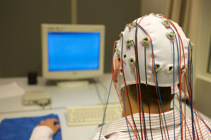
EEG (Electroencephalogram)
An EEG looks at the electrical activity in the brain
This is important in the diagnosis of epilepsy because a seizure is characterized by abnormal electrical activity in the brain
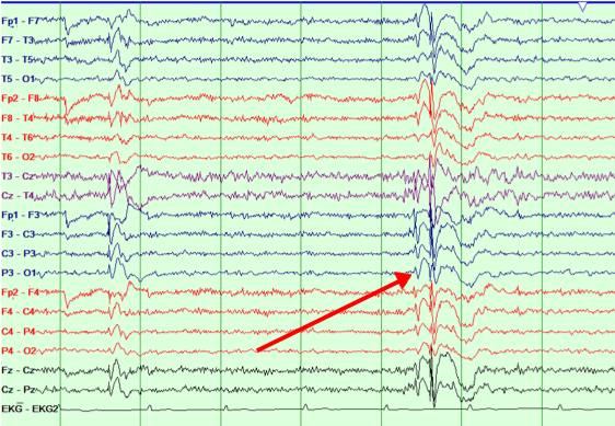
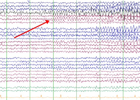
Shown below are three EEG readings:
The first image shows a page from a normal EEG
The second image shows abnormal brain waves (arrow) indicating irritability in the neurons that may cause seizures. These are called epileptiform discharges. They suggest areas where seizures may come from, or indicate a high risk of having seizures
The third image shows the seizure starting in the top leads (arrow). This is a confirmed seizure
EMU (Epilepsy Monitoring Unit)
The Epilepsy Monitoring Unit (EMU) is used for patients for two main purposes:
Diagnosis of epilepsy type.
Localization of seizures in patients with intractable epilepsy who may benefit from epilepsy surgery, vagus nerve stimulator, or responsive neurostimulation.
The EMU is a 24-hour supervised ward in the hospital where patients are voluntarily admitted to record their seizures. They may stay for up to 1-2 weeks to record enough seizures where the epileptologist is confident of seizure localization. Anti-epileptic medications may be discontinued to provoke seizures. The nurses in the ward are trained to protect and treat patients when they have seizures.
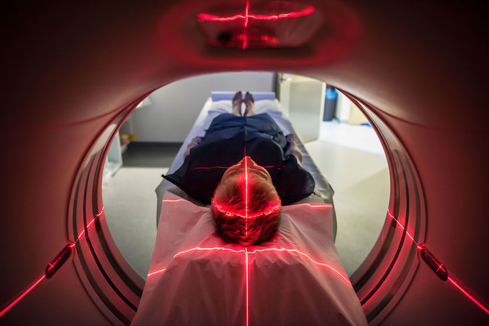
A CT scan uses low-dose X-rays to “see” the brain on a computer monitor.
The CT scanner takes pictures as taking “slices” of pictures like a loaf of bread to see inside.
This type of imaging is helpful to see if a seizure cause (for example, a tumor or a bleed in the brain) needs to be treated surgically and right away.
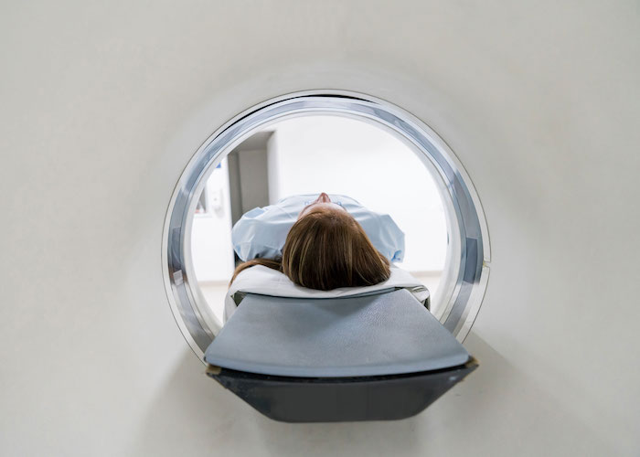
MRI (Magnetic Resonance Imaging)
An MRI is used to look for lesions in the brain tissue that may cause seizures.
In comparison to a CT scanner, the MRI can look at more clear pictures of the brain tissue and identify very subtle changes.
Sometimes, the MRI does not reveal anything. These “non-lesional” epilepsy cases range from 20-40% at epilepsy centers.
Fifty percent of these non-lesional cases can still show brain abnormalities under a microscope.
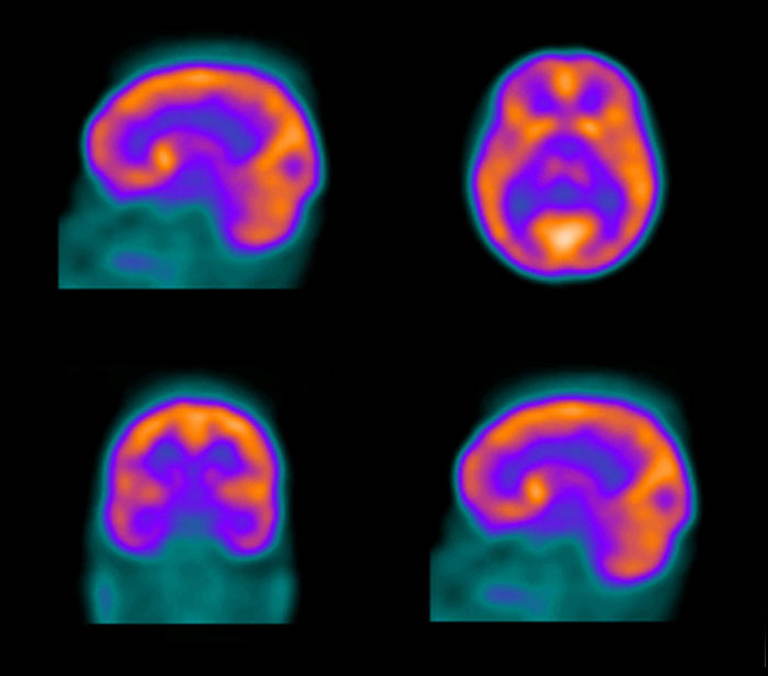
PET (Positron Emission Tomography)
A PET scan looks at brain function and may identify the region where the seizure is coming from. This may be helpful when the Brain MRI is normal or "non-lesional."
A PET scan is done by injecting a tracer into the patient’s vein to make an image which can locate the area of the brain causing seizures.
A PET scan can help localize the seizure onset zone and may help in planning for epilepsy surgery.
A single photon emission computer tomography (SPECT) scanner uses very small amounts of radioactive material to look at blood flow in the brain.
A typical result of a SPECT scan shows increased blood flow to the brain in the area where the seizure is taking place - if the scan is performed during a seizure.
The SPECT scanner will show decreased blood flow in certain lobes of the brain between seizures.
SPECT imaging can also help localize the seizure onset zone in planning for epilepsy surgery.
Magnetoencephalography (MEG)
The MEG scanner (Imagnetoencephalography scanner for human brain imaging) is a non-invasive way to measure brain activity using a very strong magnet. This can help localize areas of irritability in the brain.
MEG works by detecting the tiny (femtotesla) magnetic fluctuations at the surface of the head that arise from the brain’s electrical activity. Unlike MRI, MEG can measure the timing of brain activity with millisecond precision, allowing researchers to study the rapid brain events that underlie human cognition.
The WADA test was named after Juhn Wada, MD. It is also called the intracarotid amobarbital procedure.
This test is done to determine which hemisphere is dominant (language) and how memory is stored in each temporal lobe.
A wire, fed through the femoral artery in the upper thigh, injects aminobarbital into each carotid artery. A neuropsychologist or epileptologist will then perform memory and language testing on each side.
 There are various diagnostic tools that can help your doctor learn about your type of seizures and where they may be coming from.
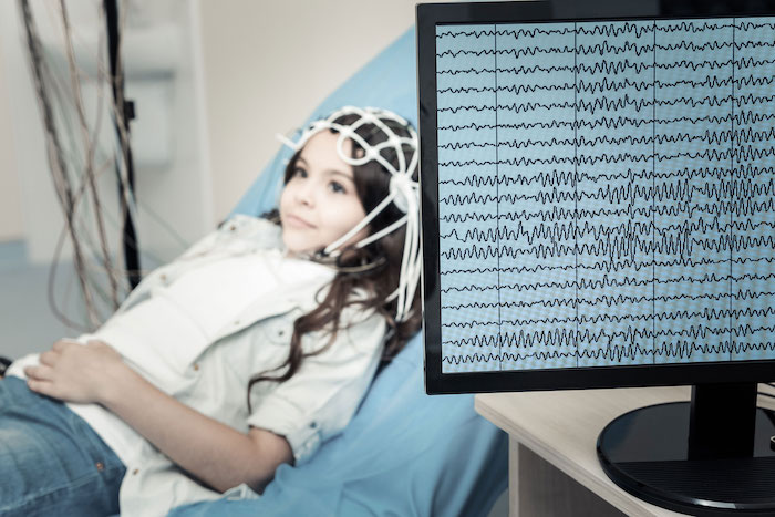 An EEG looks at the electrical activity in the brain
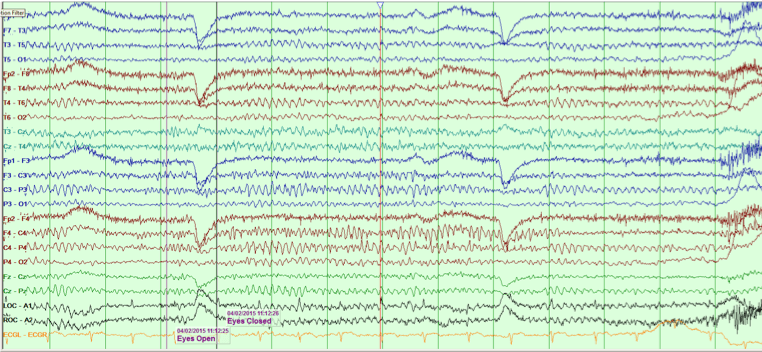
 The Epilepsy Monitoring Unit (EMU) is used for patients for two main purposes:
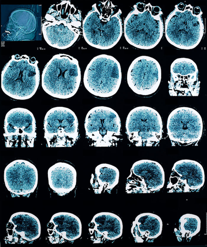
 The MEG scanner (Imagnetoencephalography scanner for human brain imaging) is a non-invasive way to measure brain activity using a very strong magnet. This can help localize areas of irritability in the brain.